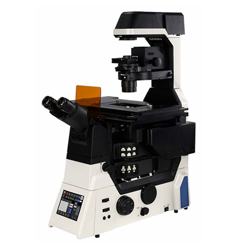
NIB1000 inverted research microscope
- Category: Biological microscopes
- Brand:Shu Yu
- Model:NIB1000
- NIB1000 provides an advanced imaging platform with 25mm field of view, to meet experimental needs of large flux observation
- INQUIRY EMAIL
NIB1000 provides an advanced imaging platform with 25mm field of view, to meet experimental needs of
large flux observation. Equipped with high-speed full-frame CMOS cameras, it provides optimal solutions
for efficient imaging. To meet the increasing demand of life science research on tissues, organs, and living
model organisms to obtain important biological data after analysis.
Fulll-motorized NIB1000-AT and coded NIB1000-M realize integration of multimodal imaging technology such
as bright field,fluorescence,DIC, phase contrast and so on, both models can choose single or double layer
optical path,anti-focus shift module can be optional which is more suitable for long-time dynamic imaging of
living cells. NIB1000 provides stable and reliable platform for confocal, superresolution, single molecule and
other advanced microscopic imaging technology.
In addition, the touch screen could achieve smooth control of electric components while recording status of
each sensor, recording user usage habits, switch application scene by one-click. Combined with powerful
acquisition and analysis software, NIB1000 is full of stability and scalability, meet a variety of experimental
needs,provide reliable, repeatable data.
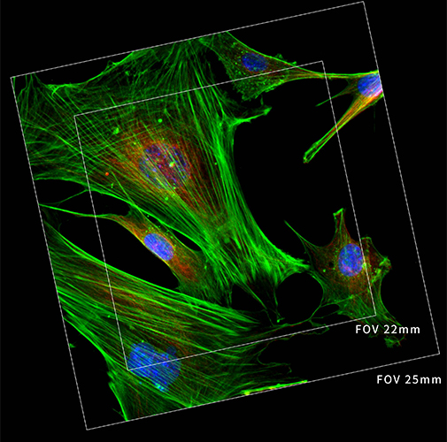
Incomparable 25mm field of view
With the research trend towards large-scale, high-flux and intelligent solutions, demand for faster data
acquisition and higher throughput is increasing day by day.No matter it is bright field imaging or fluorescence
imaging,NIB1000 large-area illuminator and large-field fluorescence attachment provides uniform and bright
cell imaging. Large size sensors and imaging interfaces that truly maximize performance of large format
detector and provide perfect imaging platform for the future as camera technology continues to evolve
rapidly.
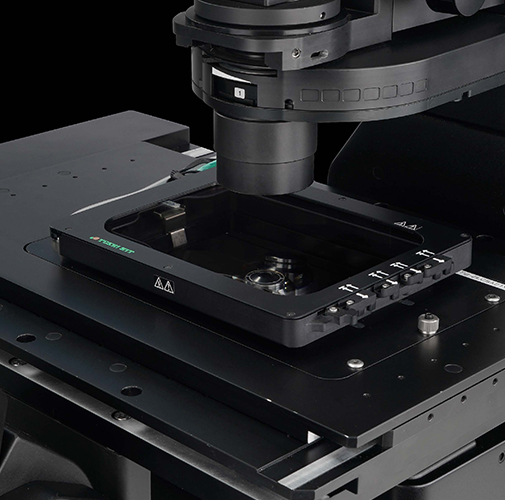
In imaging processes that require very high focusing accuracy, small temperature differences and vibrations
in the environment can significantly affect the stability of the focus. Thus, the NIB1000 is equipped with
an advanced Adaptive focusing system (AFS) that corrects and eliminates any focus drift in real time
through sophisticated static and dynamic monitoring, ensuring that the image remains sharp for long-term
observation. With the high-brightness LED fluorescent light source and live cell culture system ,NIB1000
is able to achieve multi-day time-lapse observation of live cells, making it an ideal imaging tool for more
Intuitive and ergonomic control operation
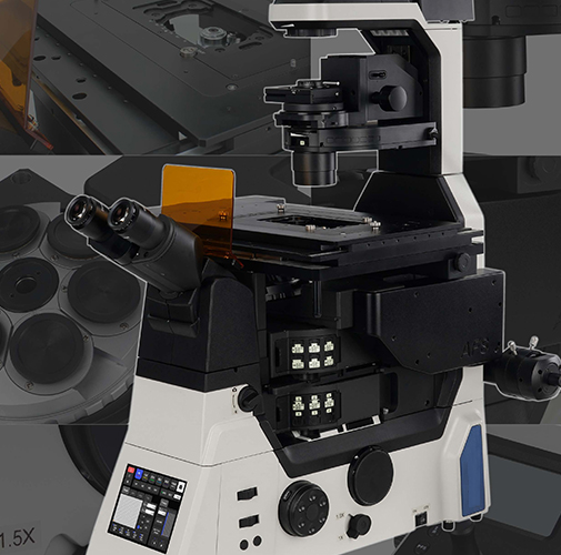
NIB1000 microscopic imaging system integrates advanced human-computer interaction
technology to give users unprecedented fine control experience. Its intuitive, simple
interface simplifies complex imaging processes, allowing researchers to navigate
complex experiments efficiently and comfortably, and significantly reducing work fatigue.
At the same time, to minimize the interference and damage to the sample, to ensure the
acquisition of the most real and complete cell dynamic information and provide strong
support for life science research.
specification
|
Item |
Specification |
|
Optical system |
NIS60 infinite optical system |
|
Eyepiece |
10×(22), with diopter adjustment -5 ~ +5; 10×(25), with diopter adjustment -5 ~ +5 |
|
viewing tube |
Seidentopf binocular observation tube, 10-40 degree tilt, pupil distance 47-78mm, eyepiece interface Φ30 |
|
viewing tube base |
eyepiece/camera(100/0,0/100),status inspection; eyepiece/camera(1000/0,0/100),external phase contras |
|
Relay lens |
0.5X, 0.7X, 1X |
|
Objective |
S-APO phase contrast objective, APO objective |
|
Nosepiece |
Code six-hole nosepiece(expansion slot), M250.75Motorized six-hole nosepiece(expansion slot), M250.75Motorized six-hole nosepiece(expansion slot), M25*0.75;AFS module included |
|
Motorized stage |
Electric control (grating type) : travel range 130 mm x 100 mm (table 445 mm x 300 mm) Maximum speed: 25mm/s; Min. : 0.1μm - Repetition accuracy: 0.5μm, can be equipped with multi-well plate, 35mm petri dish and slice three special sample damp adapter; Well Clamper is optional; |
|
Manual stage |
Three-layer mechanical moving stage, stage size : 340X230mm, moving range 130×85mm, |
|
Focusing system |
Electric control drive, coarse fine tuning 3-gear switch ((2um\80um\800um), stroke: 8.5mm above the focus and 1.5mm below; Minimum step 0.01umElectric control drive, coarse fine tuning 3-gear switch ((2um\80um\800um), stroke: 8.5mm above the focus and 1.5mm below; Minimum step 0.01um, repeat accuracy 0.1um(grating type) |
|
Touch LCD |
In front of main body, display control light source intensity, objective magnification, fluorescence band, intermediate magnification,tumable position, bertrand lens etc.; |
|
Intermediate magnification switching |
Manual 1 x, 1.5 x switching |
|
Bertrand lens |
Manual switch in and out of light path, focus adjustable |
|
Port |
Electric switching spitting ratio: Left side: visual = 100:0; Right side: Visual = 100:0; Bottom: Visual = 100:0; |
|
Illumination system |
Transmitted kohler illumination, 3W LED illumination; Field of view/aperture diaphragm; bend arm can tilt 25°; Electric optical brake optional.EPI illumination: LED optical fiber(wavelength:485nm,525nm,365nm,405nm); Field of view/aperture diaphragm; 2 hole filter insert plate; Lamp source controller; Electric brake (software control) optional. |
|
Condenser |
7-hole electric turntable for phase contrast, Hoffman phase contrast, DIC, ND filter, condenser NA=0.52, WD=30mm7-hole manual turntable, condition detection, for phase contrast, Hoffmann phase contrast, DIC, ND filter, condenser NA=0.52, WD=30mm |
|
Fluorescent turntable |
6-hole electric fluorescent turntable (B, G, U, R standard); φ25/30 light pass; Electric optical brake;6-hole manual fluorescent turntable (B, G, U, R standard); φ25/30 light pass; Manual optical brake; |
|
Double-layer fluorescence heighter |
Z-axis heightening base; Fluorescent turntable heightening seat; stage heightening seat |
|
DIC attachment |
DIC prism insert can be placed in nosepiece slot; DIC polarizer, analyzer slide plate; |
|
Controller |
XYZ electric control; Display objective magnification, fluorescence band and other states; Shortcut key setting; |
|
Software |
NOMS Pro X image analysis software |
|
Host side port |
1×M52 relay lens; Confocall left port -1.8x M52 camera relay lens |
INQUIRY


CATEGORIES
LATEST NEWS
CONTACT US
Name: Anna Zhao
Mobile:+86 15324910590
Tel:+86 15324910590
Whatsapp:+86-13526736193
Email:Henanshuyu123456@outlook.com
Add:Xiyue City, No. 33 Jinshuo Road, Hi-Tech Industrial Development Zone, Zhengzhou City, Henan Province, China